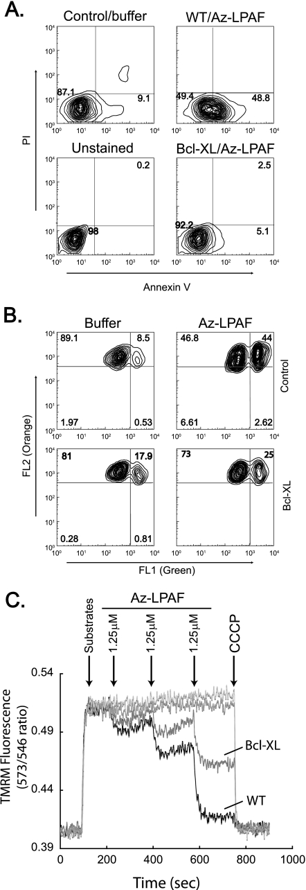
FIGURE 6.
The anti-apoptotic protein Bcl-XL protects cells and isolated mitochondria from Az-LPAF-induced depolarization and apoptosis. A, Bcl-XL suppresses Az-LPAF-induced surface expression of phosphatidyl-serine. HL-60 cells stably transfected with human Bcl-XL or empty vector were treated with 5 μm Az-LPAF or buffer for 6 h as described in the legend to Fig. 1. The cells were then stained with Alexa 488-conjugated annexin V to detect externalized phosphatidylserine and propidium iodide before cellular fluorescence was quantified by flow cytometry also as described in the legend to Fig. 1 (n = 3). B, Bcl-XL suppressed Az-LPAF-induced depolarization of mitochondria within HL-60 cells. HL-60 cells stably transfected with human Bcl-XL or empty vector were incubated with 5 μm Az-LPAF for 4 h, and then loaded with the dye JC-1 for 30 min before monomeric fluorescence in FL1 (x axis) and mitochondria internalized dye was assessed in FL2 (y axis) (n = 3). C, Bcl-XL protects isolated mitochondria from Az-LPAF-induced depolarization. Mitochondria isolated from HL-60 cells expressing Bcl-XL or empty vector were labeled with TMRM as described in the legend to Fig. 2 and given glutamate and malate at the times shown by the first arrow. Az-LPAF was then added in 1.25 μm increments at the times shown by subsequent arrows, and then the mitochondria were completely depolarized by the addition of CCCP (n = 3).

