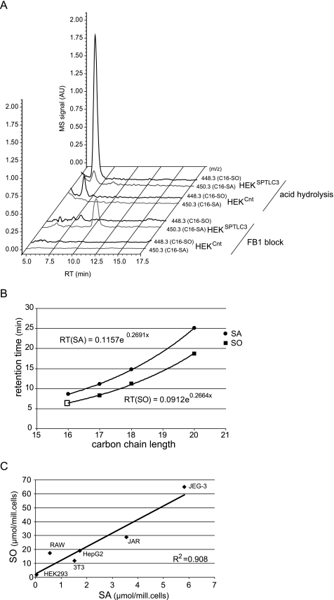
FIGURE 5.
A, identification of C16-SO in total lipid extract of HEKSPTLC3 cells. The diagram shows the single ion chromatograms for C16-SA (450.3) and C16-SO (448.3) in FB1-blocked and acid/base-treated lipid extracts from HEKCnt and HEKSPTLC3 cells. The FB1-treated HEKSPTLC3 cells showed a significant accumulation of C16-SA (450.3) which was not seen in the HEKCnt cells. No C16-SA was observed in the acid/base-treated lipid extracts from HEKSPTLC3 or HEKCnt cells. In its place a conspicuous peak appeared with the mass of C16-SO (m/z 448.3) and a retention time of 6.5 min. This peak was significantly higher in HEKSPTLC3 than in HEKCnt cells. AU, arbitrary units. RT, retention time. B, functional relationship between retention time and carbon chain length of sphingoid bases. SA and SO standards with various carbon chain lengths were separated by HPLC. The dihydro form (SA) generally eluted later than the corresponding SO form. The retention times showed a logarithmic relationship to the carbon chain length of the sphingoid bases. Based on a logarithmic regression analysis, a theoretical retention time of 6.45 min was calculated for C16-SO, which is in close concordance with the observed retention time for the C16-SO peak (6.5 min). C, C16-SA and C16-SO levels in acid/base-treated lipid extracts from various human and murine cell lines. The C16-SO levels showed a good correlation to the precursor C16-SA (R2 = 0.908).

