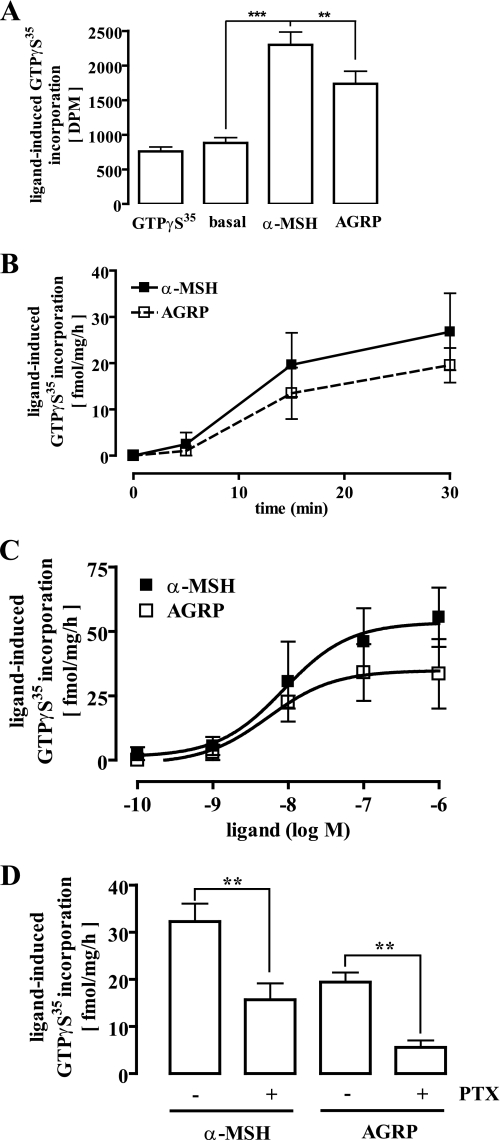
FIGURE 6.
Direct activation of Gi/o proteins by AGRP in hypothalamic GT1-7 cells. 40 μg of total membrane fractions derived from GT1-7 cells were incubated with 0. 1 nm non-hydrolysable GTPγ35S for 30 min at 30 °C. A, membranes were incubated with either 10 μm GTPγS, 1 μm α-MSH, or 100 nm AGRP. Incorporated GTPγ35S is given as total dpm. Data from one representative experiment performed in triplicate are shown. Asterisks indicate a significant (**, p < 0.01; ***, p < 0.005) difference between ligand-treated and non-treated cells. B, membranes were incubated with either 1 μm α-MSH or 100 nm AGRP for 5, 15, or 30 min. Results are expressed as the mean ± S.D. of two independent experiments carried out in duplicate. C, membranes were incubated with increasing concentrations of either α-MSH or AGRP for 30 min. Results are expressed as the mean ± S.D. of two independent experiments carried out in quadruplicate. D, before the membrane preparation cells were treated or not with 50 ng/ml PTX for 16–24 h at 37 °C. Incorporated GTPγ35S of membranes stimulated with 1 μm α-MSH or 100 nm AGRP is given as fmol/mg/h. Results are expressed as the mean ± S.E. of five independent experiments performed in quadruplicate. Asterisks indicate a significant (**, p < 0.01) difference between PTX-treated and non-treated cells.

