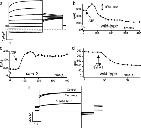
FIGURE 1.
Cytosolic ATP regulates AtCLCa currents in a reversible manner. a, AtCLCa currents recorded in a wild type vacuole in response to 5-s pulses from −77 to +83 mV in +20-mV increments followed by a 3-s tail to +33 mV. b, steady state currents measured every 10 s at +43 mV in a wild type vacuole; the arrow indicates the addition of ATP. The addition of 5 mm Mg-ATP to a wild type vacuole resulted in an initial transient increase (indicated by the double arrow), possibly because of H+-ATPase activation, followed by a current decay. c, the same experiment was conducted on vacuoles from the clca-2 knock-out mutant, where only the increase of current due H+-ATPase activation could be observed. d, 5 mm Mg-ATP was added to a wild type vacuole in the presence of the H+-ATPase inhibitor BafA1 (100 nm). e, current recordings obtained in control conditions after the addition of 5 mm Mg-ATP at the cytosolic side of the membrane and after recovery by 3 min of perfusion to wash away ATP. Membrane potential stimulation were applied every 10 s as follows; holding at 0 mV, a first pulse at +43 mV for 5 s, and a tail pulse to −33 mV for 1 s. The dashed line represents zero current level.

