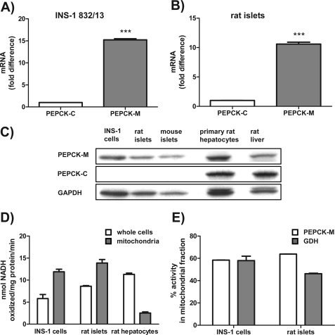
FIGURE 2.
PEPCK-M but not PEPCK-C is present in INS-1 832/13 cells, rat islets, and mouse islets. A and B, PEPCK-M mRNA expression in comparison to PEPCK-C mRNA expression by quantitative PCR in INS-1 832/13 cells (A) and rat islets (B). C, Western blots using an antibody specific for PEPCK-C and a peptide antibody specific for PEPCK-M in INS-1 832/13 cells, rat, and mouse islets. D, PEPCK activity in whole cells as well as isolated mitochondria of INS-1 832/13 cells, rat islets, and hepatocytes using a traditional method measuring NADH oxidation. E, PEPCK activity in the mitochondrial fraction of INS-1 832/13 cells and rat islets compared with glutamate dehydrogenase (GDH) activity. Error bars are S.E. and significance by t test; ***, p < 0.001.

