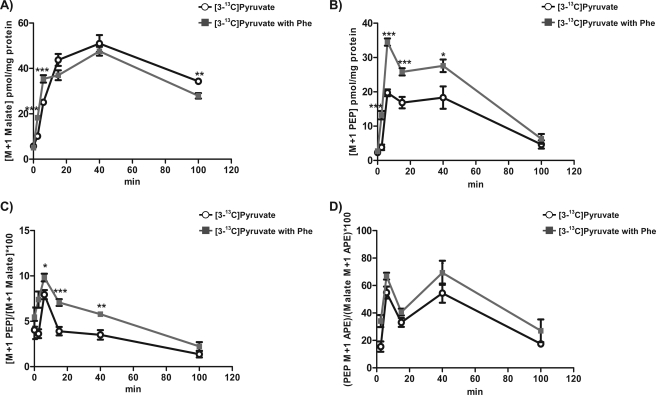
FIGURE 4.
Inhibition of pyruvate kinase by phenylalanine in INS-1 832/13 cells. A–D, INS-1 832/13 cells were preincubated in DMEM base without glucose for 60 min. Then the medium was changed to DMEM base with 5 mm [3-13C]pyruvate with (gray squares) or without (black circles) 5 mm phenylalanine and quenched at the indicated times. The concentration of 13C-labeled M+1 isotopologue of malate (A) and PEP (B) with PK inhibition. C, the ratio of [PEP M+1] concentration to [Malate M+1] concentration. D, ratio of PEP M+1 APE to malate M+1 APE (ΦPEPCK-M). Error bars are S.E. and significance was by ANOVA; *, p < 0.05; **, p < 0.01; ***, p < 0.001.

