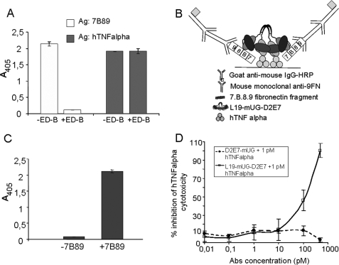
FIGURE 5.
A, ELISA using 0.8 nm D2E7-UG-L19 on the ED-B of FN (white bars) or TNF-α (dark gray bars) as antigen (Ag). The reaction was performed with or without 100 μm ED-B. B, schematic drawing; C, results of ELISA performed using TNF-α as antigen and L19-UG-D2E7 as primary antibody; after removing the excess antibody the FN fragment 7B89 was added and was detected using an antibody specific for the FN repeat 9. D, L19-mUG-D2E7 bound to the ED-B neutralizes TNF-α cytotoxicity (in situ neutralization). The cytotoxic activity of 1 pm human TNF-α was evaluated on LM cells using plates pre-coated with 7B89 and preincubated with different concentrations (0.01–500 pm) of L19-mUG-D2E7 (□) or D2E7-mUG (●). After removing unbound antibodies, hTNF-α was inhibited only by L19-mUG-D2E7, because it bound to the ED-B of FN with which the wells were pre-coated. Abs, antibodies.

