FIGURE 4.
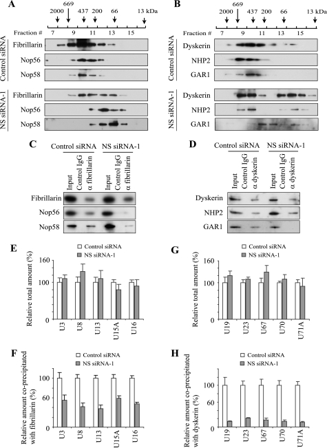
Disruption of snoRNPs by NS knockdown. A and B, Western blotting of fractions obtained from Superdex 200 gel filtration. Protein components of C/D snoRNPs were detected in A and those of H/ACA snoRNPs in B. Whole cell extract prepared from HeLa cells transfected with NS siRNA-1 or control siRNA was applied. Elution peaks of the standard marker proteins are indicated at the top. C and D, Western blotting of co-immunoprecipitated proteins with anti-fibrillarin antibody (C) and those with anti-dyskerin antibody (D) from HeLa cell extract after siRNA transfection. Input wells contain 10% of the extract used for immunoprecipitation. E and G, relative total amounts of snoRNAs in HeLa cells transfected with NS siRNA-1 in comparison to the amount in HeLa cells transfected with control siRNA. The amounts in the control cells were defined as 100%. E shows C/D snoRNAs, and G shows H/ACA snoRNAs. Mean + S.D. obtained from three independent experiments is shown. F and H, relative amounts of snoRNAs co-immunoprecipitated with anti-fibrillarin antibody (F) and those with anti-dyskerin antibody (H) from HeLa cells after transfection with NS siRNA-1.

