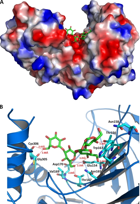
FIGURE 4.
A, molecular surface of the LPHase·laminaritetraose complex. This figure was prepared with the program PyMol. B, binding pocket of the LPHase·laminaritetraose complex. The bound laminaritetraose is drawn as green sticks, whereas ligand-binding residues of LPHase are shown as cyan sticks. Oxygen, nitrogen, and sulfur atoms are colored red, blue, and yellow, respectively.

