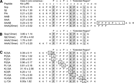
FIGURE 2.
In vitro binding analysis of AbpSH3 interactions. A, binding of the AbpSH3 to biologically relevant extended peptides. Conserved positions in AbpSH3 binding sites are highlighted in gray. Above the sequences the standard nomenclature for class II SH3 domain target sequences is defined as well as the extended region of the peptide. The residues boxed in the ArkAB sequence indicate the sequence deleted for the ΔArkB sequence used in Fig. 4. B, binding of the AbpSH3 to truncated peptide targets. C, binding of the AbpSH3 to mutant ArkA sites. The mutated sites are boxed.

