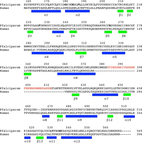
FIGURE 2.
Sequence alignment of P. falciparum and human DHODH. Secondary structure elements are defined based on the PfDHODH-DSM1 structure: α-helices are indicated by blue bars, and β-strands are indicated by green bars. Residues within 4 Å of DSM1 are displayed in bold type. The truncated surface loop in the PfDHODHΔ384–413 construct used for structure determination is shown in red.

