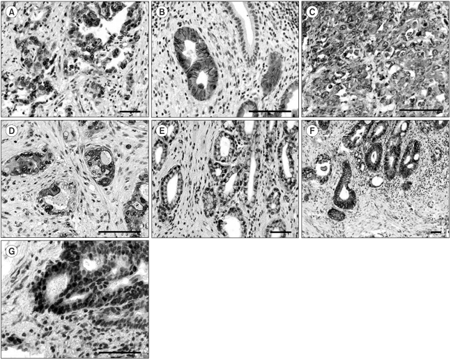
Fig. 1.
Representative immunostaining for EGFR, c-erbB2, Cox-2, c-Met and β-catenin in gallbladder cancer. A-B) Cancer cells showed selective membrane staining for EGFR (A, ×200, scale bar 100 µm) and c-erbB2 (B, ×400, scale bar 100 µm). Note the absence of immunoreactivity in normal mucosal cells. Immunohistochemistry for c-Met (C, ×400, scale bar 100 µm) and Cox-2 (D, ×400, scale bar 100 µm) displayed cytoplasmic and membrane positivity. E-G) β-catenin staining showed normal mucosal cells, with membrane staining (E, ×200, scale bar 100 µm), cytoplasmic immunoreactivity in cancer cells (F, ×100, scale bar 100 µm), and nuclear localization in cancer cells at the invasive front (G, ×400, scale bar 100 µm).

