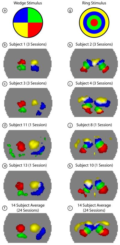
Figure 5. Activations due to phase-encoded retinotopic stimuli in multiple subjects.
(a) Legend showing the color-coding of different visual quadrants roughly corresponding to the four frames chosen from the full movie in Fig. 3. (b) Overlay of four activations (with a threshold at the half-maximum contrast of each frame) from subject 1 (all sessions averaged). Note the ability to clearly discriminate multiple visual angles within a single hemisphere. (c-e) Similar overlay images showing four visual quadrants in additional subjects. (f) Four visual quadrants shown in the average of all 24 sessions from 14 subjects. (g) Legend showing the color-coding of different visual eccentricities roughly corresponding to the four frames chosen from the full movie in Fig. 4. (h) Overlay of four activations (with a threshold at half-maximum contrast; due to uneven sensitivity to the two hemispheres, the right and left halves of the pad have different thresholds) from subject 2 (all sessions averaged). Note the ability to clearly discriminate multiple eccentricities within a single hemisphere. (i-k) Similar overlay images showing four eccentricities in additional subjects. Occasionally, the peripheral visual field (yellow) is seen only with low signal-to-noise. (f) Four visual eccentricities shown in the average of all 24 sessions from 14 subjects.

