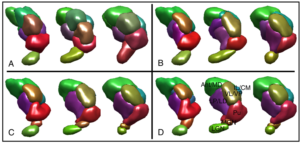
Fig. 2.
Segmentation results from three subjects’ left thalamic hemispheres are shown. Colors indicate the mean diffusion orientation in each cluster. (A) Segmentations obtained using k-means have an ellipsoidal bias and they do not correspond well between subjects. (B) Segmentations obtained using CC with no prior information are consistent among subjects. (C) Segmentation using CC with prior information are both consistent among subjects and match well with the expert segmentations. (D) Expert labeled thalami are shown. Note that even though the segmentations in (C) and (D) look very similar, they are not exactly the same (see Figure 3).

