
FIG. 4.
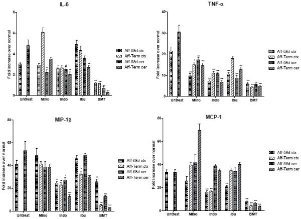
Expression of IL-6, TNF-α, MIP-1β and MCP-1 in the cortex (ctx) and cerebellum (cer) of affected trs mice (Aff) treated with minocycline (Mino), indomethacin (Indo), ibuprofen (Ibu) and BMT at PND 58 (58d) and terminal stage (Term) compared to untreated mice (Untreat) at PND 58. For the untreated mice, PND 58 corresponds to the terminal stage, for minocycline and ibuprofen the terminal stage corresponds to an average age of 70 days, for indomethacin of 67 days and for BMT-treated mice of 228 days. Each time point represent the mean ± SEM fold increase in copies of specific mRNA in the affected treated or untreated mice over the normal mice. For the pharmacological treatments five mice were used for time point, for BMT treatment 3 to 4 mice were used. Statistically significant decreases in expression levels between treated mice and untreated mice were determined by the Mann-Whitney U test (*, P 0.01-0.05; **, P 0.001-0.01; ***, P < 0.001).