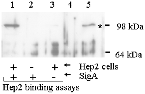
Figure 1. SigA binds specifically to HEp-2 cells.
HEp-2 cells were incubated in the presence (lane 1) or absence (lane 3) of SigA and washed to remove unbound SigA. Bound SigA was recovered by treatment of HEp-2 cells with protein sample buffer and assayed by Western analysis with anti-SigA antiserum. A control for non-specific binding of SigA to the tissue culture plate was also included (lane 2). Culture supernatants of E. coli either not expressing (lane 4) or expressing (lane 5) SigA, indicate the position of SigA on the gel (asterisk). The positions of standard molecular mass markers (kDa) are shown on the right.

