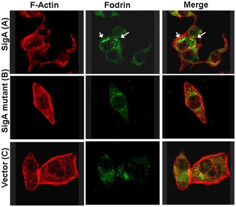
Figure 3. Effects of SigA on fodrin redistribution in epithelial cells.
HEp-2 cells were treated for 3 h with supernatant from SigA clone (A), or ΔSigA (B) or vector only used as a control (C). Cells were then fixed and stained with rhodamine-phalloidin, and anti-α-fodrin antibodies, and a fluorescein-labeled secondary anti-goat antibody. Intracellular aggregates of fodrin are indicated by arrows.

