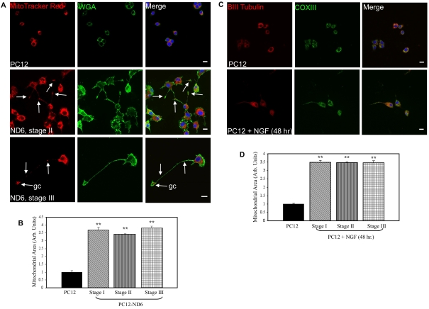
Figure 3. PC12-ND6 cells display increased mitochondrial area.
(A) Staining of the mitochondrial compartment. PC12 and PC12-ND6 cells were stained with MTR (red) and the plasma membrane marker Alexa Fluor® 488-conjugated WGA (green) and TO-PRO®-3 (blue). White arrows indicate the presence of mitochondria within neurites or growth cones (gc). Scale bar, 10 μm. (B) Quantification of mitochondrial area in PC12 and PC12-ND6 cells at different stages of neuronal differentiation. Results are means±S.D. for three independent experiments, with a minimum of n = 150 cells for each cell type and a minimum of n = 50 cells per stage. **P<0.0001 (Student's t test). (C) Staining of the mitochondrial compartment in untreated and NGF-differentiated PC12 cells. Untreated and NGF-treated PC12 cells for 48 h (100 μg/ml) were stained with antibodies against β-III-tubulin (red), COX III (green) and subsequently counterstained with the TO-PRO®-3 (blue). Scale bar, 10 μm. (D) Quantification of mitochondrial area in untreated and NGF-treated PC12 cells. Results are means±S.D. for three independent experiments, with n = 150 cells per stage. **P<0.0001 (Student's t test).

