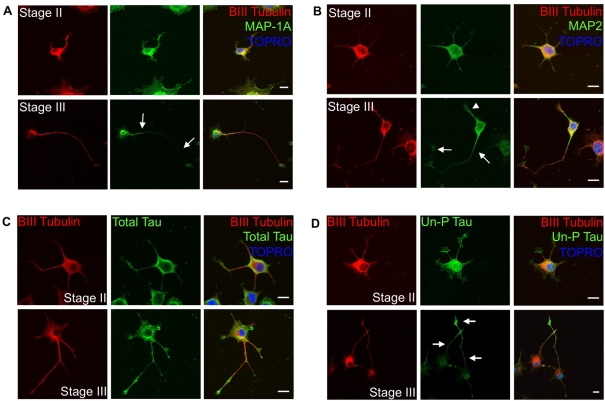
Figure 6. Reorganization of microtubules and corresponding microtubule-binding proteins in PC12-ND6 cells.
(A) Stage-specific redistribution of MAP-1A. PC12-ND6 cells were labelled with anti-β-III-tubulin antibody (red), anti-MAP-1A antibody (green) and TO-PRO®-3 (blue). In stage II PC12-ND6 cells (top row), MAP-1A is evenly distributed throughout the cell. In stage III PC12-ND6 cells (bottom row), a MAP-1A gradient has been established with MAP-1A labelling excluded from the nascent axon, as indicated by white arrows. Scale bar, 10 μm. (B) Stage-specific redistribution of MAP-2. PC12-ND6 cells were labelled with anti-β-III-tubulin antibody (red), anti-MAP-2 antibody (green) and TO-PRO®-3 (blue). In stage II PC12-ND6 cells (top row), MAP-2 is evenly distributed throughout the cell. In stage III PC12-ND6 cells (bottom row), a gradient of MAP-2 has been established, with MAP-2 labelling excluded from the growing axon (white arrows), but present within developing dendrites (white arrowhead). Scale bar, 10 μm. (C) Total tau is evenly distributed in PC12-ND6 cells at stages II and III. PC12-ND6 cells were labelled with anti-β-III-tubulin antibody (red), an antibody recognizing all forms of tau (total tau, green) and TO-PRO®-3 (blue). Scale bar, 10 μm. (D) Expression of unphosphorylated tau is enriched in the distal portion of growing axons in stage III PC12-ND6 cells. PC12-ND6 cells were labelled with anti-β-III-tubulin antibody (red), antibody recognizing only the unphosphorylated form of tau (Un-P Tau, green) and TO-PRO®-3 (blue). In stage II PC12-ND6 cells (top row), unphosphorylated tau is evenly distributed throughout the soma and neurites. In contrast, stage III PC12-ND6 cells (bottom row) display unphosphorylated tau enriched within the distal portion of the growing axon, as indicated by arrows. Scale bar, 10 μm.

