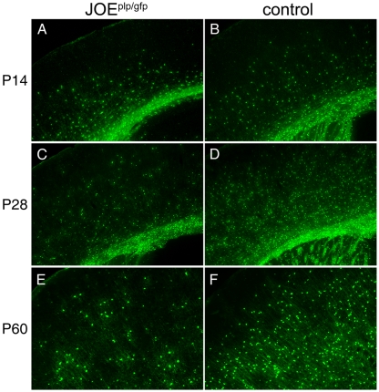
Figure 6. Fewer GFP+ OLs detected in the JOE cortex.
Coronal cortical sections from JOE×PLP–GFP (JOEplp/gfp) hemi and WT mice at P14 (A and B), P28 (C and D) and P60 (E and F). The number of GFP-labelled OLs in the JOEplp/gfp mice were reduced and localized in disorganized clusters in comparison with the more radially aligned OLs labelled in the WT mice (control). Magnification ×50 (A–D), ×100 (E and F).

