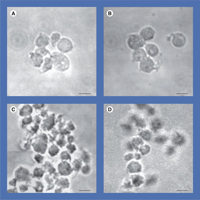
Figure 3. Differential interference contrast analysis of effects of human-derived nanoparticles on activation and aggregation of human platelets.

Representative images show unstimulated platelets (control) (A) maintained discoid morphology with moderate activation, whereas platelets coincubated with 150 nephelometric turbidity unit human-derived nanoparticles (B) exhibited a larger proportion of cells with spherical enlargement, together with shape changes and pseudopodia. Platelets exposed to thrombin receptor activator peptide (10 μM) (C) aggregated but platelets incubated with human-derived nanoparticles for 15 min prior to stimulation with thrombin receptor activator peptide (D) showed fewer and smaller aggregates. Cumulate data from these experiments are quantified in Figure 4. Scale bars represent 2 μm.
