
Fig. 4.
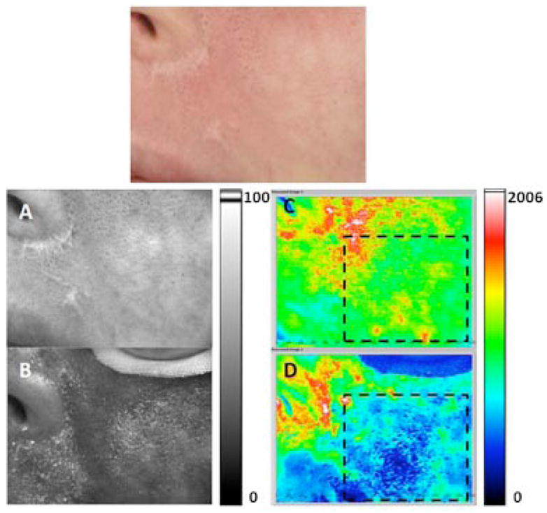
Example of moderate DoH achieved after laser therapy. Top: Photograph of user-specified region of interest (ROI) identified on a female Caucasian patient with a facial PWS birthmark. A, B: Grayscale and (C, D) corresponding SFI maps of ROI treated with a PDL. Images were acquired (A, C) immediately before, and (B, D) 40 minutes after, treatment. Areas enclosed in dashed lines in (C) and (D) were selected to calculate the change in perfusion and DoH. B perfusion was decreased by 50% after treatment, with a post-treatment DoH of 0.38 (i.e., moderate level).