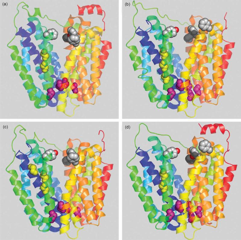
Figure 1.
Homologous bacterial MFS transporters threaded into LacY structure. Transmembrane helices are depicted as ribbons colored from blue (helix I) to red (helix XII). The cytoplasmic side is at the top. Conserved Gly residues in helices I and V are shown as yellow spheres. Conserved Gly residues on the extracellular surface between the N and C-terminal six-helix bundles are shown as pink spheres. Conserved aromatic residues facing hydrophilic cavity are shown as grey spheres. (a) LacY (PDB code 1PV7); (b) E. coli sucrose permease CscB; (c) E. coli raffinose permease RafB; (d) Bacillus megaterium fructose permease FruP.

