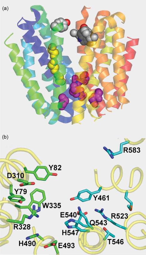
Figure 3.
Predicted structure of human FLJ20160 protein transmembrane domains threaded into LacY structure. (a) Organization of transmembrane helices with cytoplasmic side at the top. Connecting loops, and N and C termini are removed. Transmembrane helices are depicted as ribbons colored from blue (helix I) to red (helix XII). Conserved Gly residues in helices I and V are shown as yellow spheres. Conserved Gly residues on the extracellular surface between the N and C-terminal six-helix bundles are shown as pink spheres. Conserved aromatic residues facing hydrophilic cavity are shown as grey spheres. (b) Positions of residues that may be involved in sugar binding and/or H+ translocation viewed from the cytoplasmic side of the hydrophilic cavity. Amino acid residues with possible importance for sugar binding (green) or H+ translocation (cyan) are shown as sticks.

