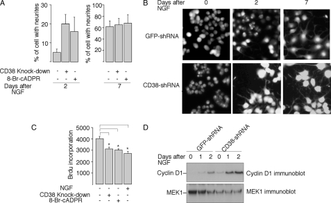
FIGURE 7.
The role of the CD38/cADPR signaling in NGF-induced neuronal differentiation. A, CD38-knockdown or treatment with 8-Br-cADPR (100 μm) accelerated NGF-induced neurite outgrowth in PC12 cells. Data are expressed as means ± S.D., n = 3. B, representative fluorescence images of Fura-2 labeled wild-type or CD38-knockdown PC12 cells, both treated with NGF (50 ng/ml) for the indicated number of days. C, inhibition of the BrdUrd incorporation into PC12 cells by 8-Br-cADPR, CD38-knockdown, or NGF. Cells were plated as described under “Experimental Procedures,” and treated with or without NGF (50 ng/ml) or 8-Br-cADPR (100 μm) for 24 h before assayed for BrdUrd incorporation. The data are expressed as means ± S.D., n = 3. The * symbols indicate the results of the Student's t test analysis, p < 0.005, compared with PC12 wild-type cells without treatment. D, expression of cyclin D1 in CD38-shRNA expressed PC12 cells, or control cells expressed GFP-shRNA, in response to NGF (50 ng/ml) treatment for the indicated times. MEK-1 immunoblot was used as the internal control.

