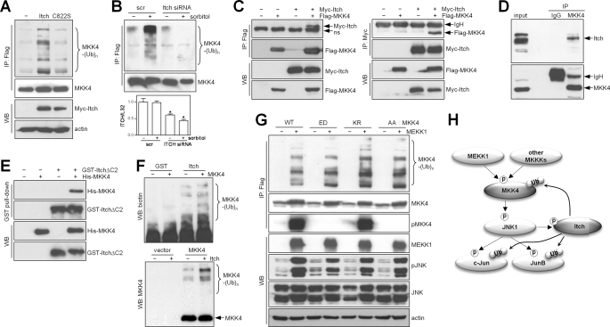
FIGURE 4.
Itch is an E3 ligase for MKK4. A, immunoprecipitation (IP)/Western blot (WB) of 293T cells transfected with wild-type Itch or an inactive E3 ligase mutant (C822S) to detect ubiquitinated MKK4. Actin was used as a loading control. B, immunoprecipitation/Western blotting to detect ubiquitinated (upper panel) or total (middle panel) MKK4 in 293T cells transfected with scrambled (scr) control or Itch small interfering RNA (siRNA), which effectively knocked down Itch on the basis of quantitative reverse transcription-PCR analysis. In the bar graph (lower panel), values were normalized on the basis of ribosomal protein L32. *, p < 0.05 scrambled versus Itch (n = three per group). C, co-immunoprecipitation of Itch and MKK4 from 293T cells cotransfected with Myc-Itch and FLAG-MKK4. Binding was analyzed by performing anti-FLAG immunoprecipitation/anti-Myc Western blotting (left panels) or anti-Myc immunoprecipitation/anti-FLAG Western blotting (right panels). Arrows point to specific bands of interest. ns, nonspecific band. D, co-immunoprecipitation of endogenous Itch and MKK4 in 293T cells by anti-MKK4 immunoprecipitation/anti-Itch Western blotting. E, in vitro binding assay using GST-ItchΔC2 and His-MKK4 proteins purified from Escherichia coli. F, in vitro ubiquitination assay with purified His-MKK4 from E. coli (upper panel) and immunoprecipitated FLAG-MKK4 from 293T cells (lower panel), GST-ItchΔC2, and UbcH6 as an E2 ubiquitin-conjugating enzyme. Ubiquitinated MKK4 was detected by Western blotting using anti-biotin (upper panel) and anti-MKK4 (lower panel) antibodies. G, immunoprecipitation/Western blotting to detect MKK4 ubiquitination (upper panel) and phosphorylated (p) and total proteins (lower panels) in 293T cells cotransfected with MEKK1 and MKK4 (wild-type (WT) or mutant). ED, constitutively active S257E/T261D mutant; KR, kinase-dead K131R mutant; AA, kinase-dead S257A/T261A mutant. H, diagrammatic illustration of a negative feedback loop involving MKK4/JNK/Itch and other substrates of Itch and JNK (c-Jun and JunB). Arrows indicate directional phosphorylation (p) and ubiquitination (Ub).

