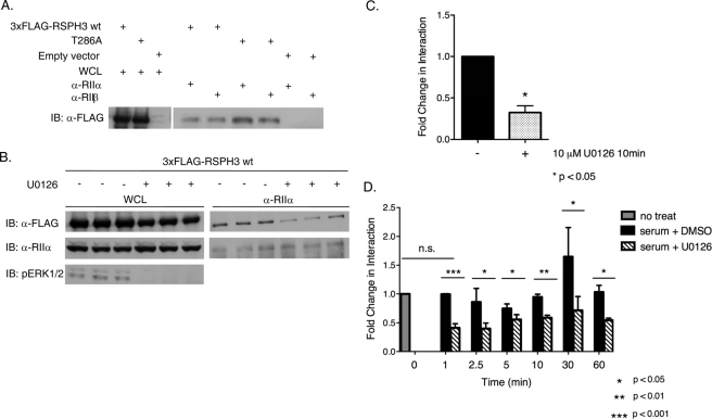
FIGURE 6.
ERK1/2 activity modulates the AKAP function of RSPH3. A, endogenous RIIα or RIIβ was immunoprecipitated from HEK293 cells expressing 3xFLAG-RSPH3 wild type (wt), T286A, or empty vector. Samples were immunoblotted (IB) with anti-FLAG antibody. Fold change in interaction (as described under “Results”) was determined using a ratiometric analysis of the intensity of the RSPH3 band relative to the whole cell lysate (WCL) for each protein. The experiment was performed three times each for RIIa and for RIIb. Statistical significance of the fold change in interaction was determined using a two-tailed t test, p value = 0.011. B, HEK293 cells expressing wild-type 3xFLAG-RSP3H were placed in serum-free medium for 12 h prior to stimulating with serum in the presence of 10 μm U0126 or diluent for 10 min. Endogenous RIIα was immunoprecipitated from the lysates, and the material was immunoblotted with the indicated antibodies. Data of B are shown in C as mean + S.E., n = 3. The fold change in interaction was based on the ratiometric analysis, and values were normalized to the DMSO-treated control. Statistical significance was analyzed using a two-tailed t test, p value = 0.0139. D, HEK293 cells expressing 3xFLAG-RSP3H were stimulated with serum with 10 μm U0126 or DMSO for the indicated times. RIIα was immunoprecipitated, and the precipitates were blotted with anti-FLAG. Fold change was determined as above. Samples were normalized to the untreated control. Data represent mean + S.E., n = 3. Statistical significance was analyzed using a two-tailed t test with p values as indicated.

