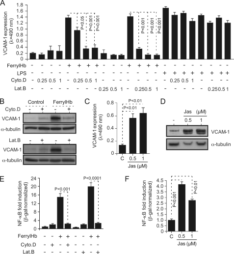
FIGURE 8.
The proinflammatory effect of ferrylHb is dependent on actin polymerization. A, confluent HUVEC were exposed to cytochalasin D (Cyto.D; 0.25–1 μm; 30 min), LatB (0.25–1 μm; 30 min), or vehicle (−) and not further treated (−) or exposed (8 h) to ferrylHb (20 μm) or LPS (100 ng/ml). VCAM-1 expression was detected by cellular ELISA. B, confluent HUVEC were treated with cytochalasin D (0.5 μm; 30 min; +), latrunculin B (0.5 μm; 30 min; +) or vehicle (−) and not further treated (Control) or exposed (8 h) to ferrylHb (20 μm). Proteins were detected in whole cell lysates by Western blotting. C and D, confluent HUVEC were exposed (8 h) to jasplakinolide (Jas) or to vehicle (C), and VCAM-1 protein expression was measured by cellular ELISA (C) or by Western blotting (D). E, confluent HUVEC were transduced (48 h) with NF-κB-Luc and LacZ recombinant adenovirus, treated with cytochalasin D (0.5 μm; 30 min; +), latrunculin B (0.5 μm; 30 min; +), or vehicle (−), and then not further treated (−) or exposed (8 h) to ferrylHb (20 μm). Luciferase was normalized to β-galactosidase units, and values are represented as mean fold-induction versus control non-treated cells ± S.D. (n = 3) from 1 of 3 independent experiments. p values were calculated with the unpaired Student's t test. F, confluent HUVEC were transduced as in E and either not treated, treated (8 h) with jasplakinolide, or vehicle (C), and the rest of the procedure was done as in E. Results in A, C, E, and F are the mean values ± S.D. (n = 3) from 1 of 3 independent experiments. p values were calculated with ANOVA and the Tukey-Kramer multiple comparison test. Immunoblots in B and D are representative of three independent experiments.

