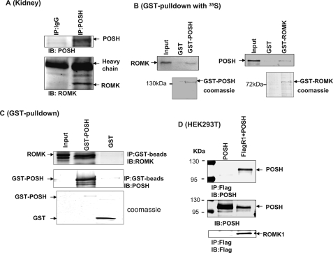
FIGURE 2.
ROMK channels are associated with POSH. A, a Western blot demonstrating that ROMK channels are immunoprecipitated with POSH in the rat kidney. B, a GST pulldown experiment shows that GST-POSH fusion protein is able to pull down ROMK1 (left panel) and that GST-ROMK protein is also able to associate with POSH (right panel). GST-POSH or GST-ROMK1 was bound to glutathione-Sepharose beads and was incubated with either 10 μl of purified ROMK1 protein or POSH protein labeled with [35S]methionine in NETN buffer. After washing, the bound proteins were resolved by 10% SDS-PAGE followed by autoradiography. C, GST pulldown experiments showing the association between GST-POSH and ROMK1. D, a Western blot showing that POSH is specifically immunoprecipitated with FLAG-tagged ROMK1 in 293T cells transfected with FLAG-R1+POSH but not in cells transfected with POSH alone. Two bands on the left side of the gel are molecular size markers. IP, immunoprecipitation; IB, immunoblot.

