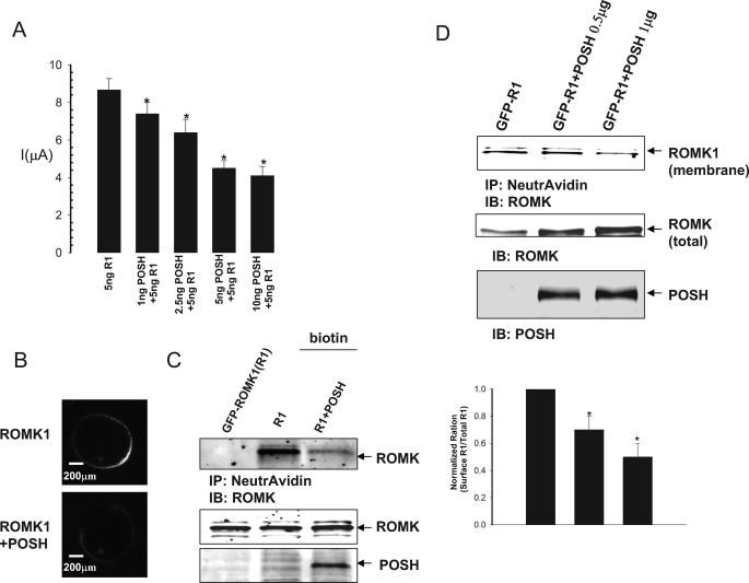
FIGURE 5.
Effect of POSH on ROMK1 channel activity and expression. A, effect of POSH on potassium current in oocytes injected with ROMK1 and POSH at different concentrations. The asterisks indicate a significant difference between control (without POSH) and POSH-injected oocytes. The experimental numbers are at least 11 for each condition. B, a confocal image showing the effect of POSH on the surface expression of GFP-ROMK1 in oocytes. C, POSH decreases the surface expression of ROMK1 channels in HEK293T cells transfected with POSH+GFP-ROMK1 or GFP-ROMK1 alone. Sulfo-NHS-SS biotin was used to label the surface ROMK1, and 50 μg of protein from cell lysates was used for the experiments. D, biotin labeling experiments showing the effect of increased POSH expression on the ROMK1 expression in plasma membranes of HEK293T cells. A bar graph summarizing the results from four experiments is shown on the bottom. IP, immunoprecipitation; IB, immunoblot.

