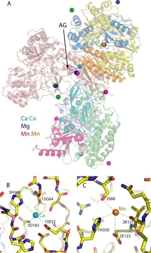
FIGURE 4.
Bound cations. A, an overview. The three structures (O4, O2, and RND) containing three different types of bound cations were superposed, and the cations are shown as spheres. Oxidized domain structure (O4) is shown in cartoon representation with subunit Nqo1 is in yellow, Nqo2 is in blue, Nqo3 is in salmon, Nqo4 is in green, Nqo5 is in violet, Nqo6 is in red, Nqo9 is in cyan, and Nqo15 is in orange. AG indicates the acidic groove. Ca2+ ions are shown in green except the tightly bound Ca2+ present in all structures, which is in cyan. Mg2+ is in blue, and Mn2+ is in magenta except the Mn2+ atom bound in the putative iron-binding channel, which is in orange. B, close-up of the tightly bound Ca2+ ion with interacting residues indicated. C, close-up of the Mn2+ ion bound in the putative iron-binding channel at the interface with frataxin-like subunit Nqo15. Residues interacting with the metal are indicated, including possible weak interaction with carbonyl of 2Lys121. The prefixes before the residue names indicate the subunit number.

