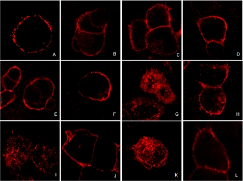
FIGURE 6.
Agonist-mediated GPR55E internalization in HEK293 cells transiently transfected with HA-GPR55E. Live HEK293 cells expressing HA-GPR55E prelabeled with anti-HA mouse antibody and Alexa Fluor 568 secondary antibody were treated with various compounds, and membrane staining was imaged by confocal microscopy at 63× magnification. Receptor internalization resulted in a loss of cell surface immunofluorescence. A, vehicle-treated cells show predominantly plasma membrane receptor staining. B–F, treatment with 30 μm 2-AG, 30 μm anandamide, 30 μm THC, 30 μm O-1602, and 10 μm CP55,940, respectively, resulted in no loss of receptor surface staining and resembled vehicle treated cells. G, I, and K, internalization of membrane-bound GPR55 occurs upon treatment with 3 μm LPI, 30 μm SR141716A, and 30 μm AM251, respectively. H, J, and L, the co-application of 3 μm CP55,940 in the presence of 3 μm LPI, 30 μm SR141716A, and 30 μm AM251, respectively, attenuated receptor internalization and restored plasma membrane receptor staining.

