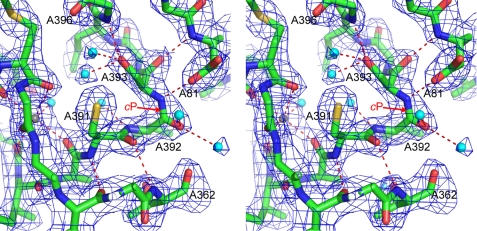
FIGURE 3.
An unusual cis-peptide bond between Val-A392 and Ser-A393. The stereo figure shows residues around the cis-peptide (labeled cP). Polar contacts between residues are shown as red dotted lines, water molecules as cyan spheres, and a metal ion as a gray sphere. The density, shown as a blue mesh, is a 2mFo − DFc map, contoured at 1.3 σ.

