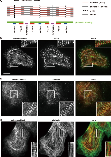
FIGURE 1.
Localization of Fhod3 in cultured rat cardiomyocytes. A, shown is a representation of the sarcomere structure (upper panel) and relative localization of Fhod3 and other sarcomeric proteins from B–D (lower panel). B–D, neonatal rat cardiomyocytes were subjected to immunofluorescent double staining for endogenous Fhod3 (red) and α-actinin (green) (B), myomesin (green) (C), or phalloidin (green) (D). For Fhod3 staining, the anti-Fhod3-(650–802) polyclonal antibodies were used. Scale bar, 10 μm.

