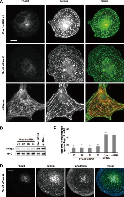
FIGURE 2.
Fhod3 knock down disrupted sarcomere assembly in rat cardiomyocytes. A, cardiomyocytes were transfected with Fhod3-specific siRNAs 2 and 3 and cultured for 48 h. Cells were fixed and double stained with the antibodies against anti-Fhod3-(650–802) (red) and α-actinin (green). Scale bar, 10 μm. Images for cells transfected with Fhod3-specific siRNAs 1 and 4 are shown in supplemental Fig. S3. B, the protein level of Fhod3 was determined by immunoblot analysis using anti-Fhod3-(873–974). As a control, a monoclonal antibody against the cardiac myosin heavy chain (MHC) was used. C, cells with well developed sarcomere organization were counted, and the percentages from four independent transfections are expressed as the means ± S.D.; cells with organized sarcomere are defined as ones displaying at least five repeated α-actinin bands with a width more than 2 μm by α-actinin staining. D, cardiomyocytes transfected with Fhod-specific siRNA 4 were fixed and stained with the antibodies against anti-Fhod3-(650–802) (red) and α-actinin (green) and with phalloidin (blue). Scale bar, 10 μm.

