FIGURE 6.
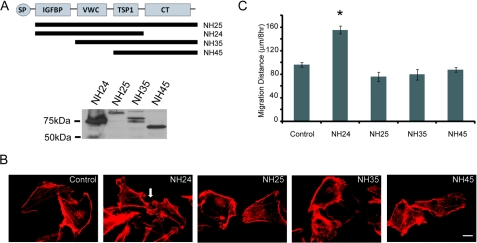
Intracellular CCN3 lacking the CT domain promotes migration. A, top panel, schematic diagram showing the CCN3 deletion mutants. Bottom panel, Western analysis with anti-GFP antibody showing the expression of recombinant GFP-CCN3 fusion proteins expressed in MDA-231 cells. B, actin cytoskeleton of CCN3 mutant-expressing cells as visualized by Alexa 568-conjugated phalloidin. White arrow indicates the membrane protrusions present in NH24-expressing cells. Scale bar, 10 μm. C, migration distance of CCN3 mutants-expressing cells compared with control as determined by wound healing assays (* = p < 0.001, one-way analysis of variance).

