FIGURE 2.
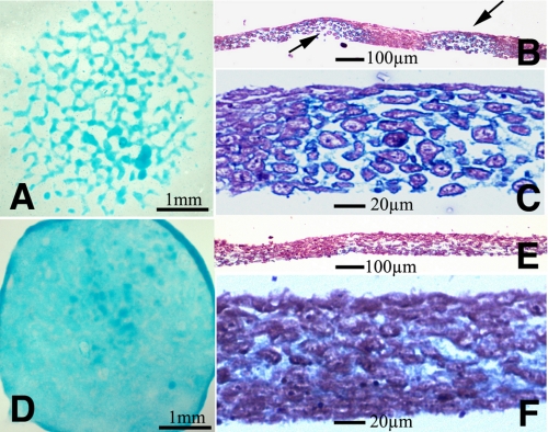
Effect of exogenous TGFβs in limb mesenchyme micromass cultures. Five-day-old micromass cultures representative of control (A–C) and cultures treated with TGFβ1 at the beginning of day 3. A shows the characteristic nodular pattern of the control cultures stained for cartilage with Alcian blue. B and C are semi-thin histological sections to show the presence of cartilage nodules in the culture. B is a low magnification view showing two cartilage nodules (arrows) separated by a fibrous like tissue. C is a detailed view of a cartilage node showing the rounded morphology of chondrocytes and the Alcian blue-positive extracellular matrix. D–F illustrate the morphology of the experimental micromasses to show the absence of cartilage nodes. Note the uniform and weak Alcian blue staining in D and the fibrous appearance of the tissue in the semi-thin sections (E and F).

