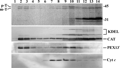
FIGURE 3.
35S-Labeled prethiolase is specifically imported into peroxisomes. A ΔC1PEX5L-supplemented PNS fraction was incubated with 35S-labeled prethiolase in import buffer containing ATP for 45 min. After trypsin treatment and inactivation of the protease, the complete import mixture was diluted with SEM buffer and subjected to Nycodenz gradient centrifugation. The gradient was then fractionated from the bottom (lane 1) to the top (lane 14), and equal aliquots from each fraction were subjected to SDS-PAGE and Western blotting. The nitrocellulose membrane was exposed to an x-ray film to detect the 35S-labeled protein (top panel) and afterward probed with the following antisera: anti-KDEL (KDEL; recognizes GRP72 and GRP98, two endoplasmic reticulum proteins), anti-cytochrome c (Cyt c; a mitochondrial marker), anti-catalase (CAT; a peroxisomal enzyme), and anti-PEX13. This last serum recognizes on trypsin-treated peroxisomes a 30-kDa fragment of PEX13 (PEX13′) (46). Note that catalase remaining at the top of the gradient (lanes 11–14) results from leakage of peroxisomes during preparation of PNS fractions. p-T and m-T, precursor and mature forms of thiolase, respectively. The numbers at the right indicate the molecular masses of the applied standards in kDa.

