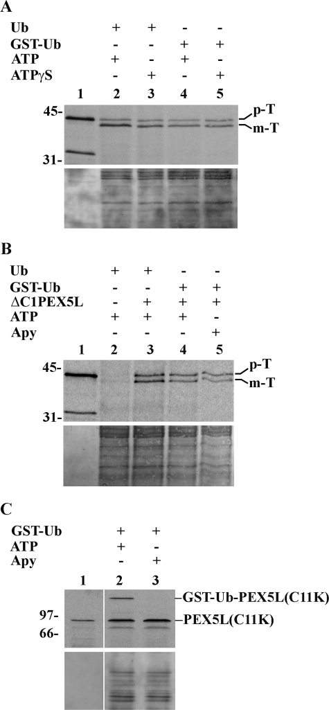
FIGURE 4.
Import of prethiolase into peroxisomes does not require hydrolysis of cytosolic ATP. A, import reactions assembled with components pretreated with 0.3 mm ATP (see “Experimental Procedures”) and supplemented with either bovine Ub or GST·Ub were performed in the presence of 3 mm ATP or 3 mm ATPγS, as indicated. After trypsin treatment, the organelles were isolated by centrifugation and analyzed as described in the legend for Fig. 2. The autoradiograph (upper panel) and the corresponding Ponceau S-stained membrane (lower panel) are shown. Lane 1, 10% of 35S-labeled prethiolase solution used in each lane. B, a rat liver PNS was pretreated with 0.3 mm ATP for 5 min and divided into four equal aliquots (lanes 2–5). The first and second aliquot (lanes 2 and 3, respectively) received bovine ubiquitin and 3 mm ATP; the third aliquot received GST·Ub and 3 mm ATP (lane 4); and the fourth received GST·Ub and apyrase (Apy). After 3 min at 37 °C, the first aliquot received 35S-labeled prethiolase preincubated with ATP in the absence of ΔC1PEX5L, whereas the second and third aliquots received 35S-labeled prethiolase preincubated with ATP in the presence of ΔC1PEX5L (lanes 3 and 4). The fourth aliquot received 35S-labeled prethiolase preincubated with ATP plus ΔC1PEX5L and treated with apyrase (see “Experimental Procedures” for details). After 45 min at 37 °C, the samples were treated with trypsin and processed as described above. Lane 1, 10% of the 35S-labeled prethiolase used in the import reactions. The autoradiograph (upper panel) and the corresponding Ponceau S-stained membrane (lower panel) are shown. p-T and m-T, precursor and mature forms of thiolase, respectively. C, in vitro assays using 35S-labeled PEX5L(C11K). The experimental conditions used in lanes 2 and 3 were exactly the ones used in lanes 4 and 5 of the experiment shown in B, respectively, with the exception that recombinant ΔC1PEX5L was omitted because it strongly competes with the radiolabeled protein for the DTM. At the end of the 45-min incubation at 37 °C, the organelles were sedimented and processed for SDS-PAGE. Note that PEX5L(C11K) is as functional as normal PEX5L in these assays (23). It was used here for practical reasons because the GST·Ub·PEX5L(C11K) conjugate, unlike the GST·Ub·PEX5L, is not destroyed by prolonged incubation in the presence of GSH and can be analyzed under normal SDS-PAGE conditions (23). The complete absence of GST·Ub·PEX5L(C11K) in lane 3 indicates that the amount of apyrase used in these assays efficiently depletes ATP from the reactions. Lane 1, 10% of 35S-labeled PEX5L(C11K) used in each reaction. Numbers to the left indicate the molecular masses of protein standards in kDa.

