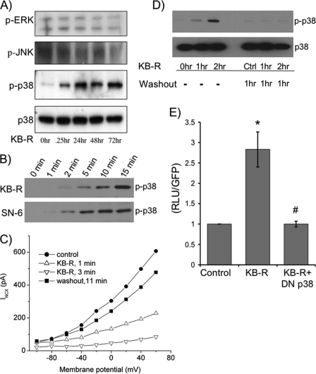
FIGURE 3.
Inhibition of NCX1 by KB-R7943 activates p38. A, lysates from cells treated with 10 μm KB-R at different time points were subjected to SDS-PAGE followed by Western blotting to determine the activation of MAPKs by probing with phosphorylation-specific antibodies to ERK (p-ERK), JNK (p-JNK), and p38 (p-p38). The data are representative of six independent experiments. B, cells were treated with KB-R or another NCX1 inhibitor, SN-6, for 1–15 min. Cells were lysed and subjected to SDS-PAGE followed by Western blotting to determine the activation by phosphorylation of p38 (p-p38). The Western blot is representative of four experiments. C, representative current-voltage relationship for the exchanger 1 min before application of KB-R (filled circles), 1 min after the application of 10 μm KB-R (open triangles), 3 min after KB-R (inverted open triangles), and 11 min after beginning washout (filled squares). The data are representative of five experiments. D, cardiomyocytes were treated with 10 μm KB-R for 1 and 2 h and washed out for 1 h, and then cells were lysed and subjected to SDS-PAGE followed by Western blotting to determine the activation of p38. Ctrl, control. E, isolated adult cardiomyocytes were sequentially co-infected with the 1831Ncx1 promoter luciferase reporter adenovirus (m.o.i. of 1.5) followed after 8 h with the dominant negative p38 adenovirus. Cells were treated with or without 10 μm KB-R for 48 h. Cells were lysed in reporter buffer and luciferase activity was determined relative to GFP levels. *, p < 0.05 when compared with control; #, p < 0.05 when compared with KB-R stimulated cells as determined by ANOVA (n = 6).

