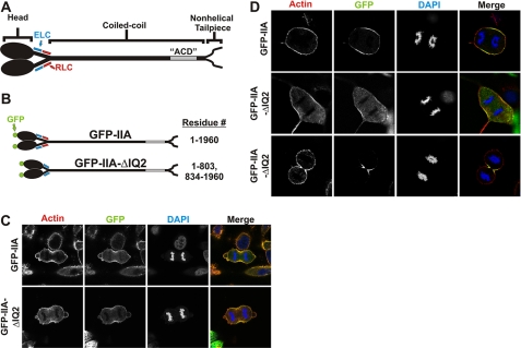
FIGURE 1.
RLC-independent localization of myosin to furrow in HeLa and COS-7 cells. A, diagram of myosin IIA. GFP was conjugated to the amino terminus of the MHC. B, diagram of GFP-IIA constructs. GFP-IIA-ΔIQ2 removes the RLC binding site known as the IQ2 motif. C and D, at 72 h after transfection, HeLa (C) or COS-7 (D) cells expressing GFP-IIA (top row) or GFP-IIA-ΔIQ2 in early anaphase (middle row) or late anaphase (lower row) were fixed and stained with phalloidin-568 (red) for actin and DAPI (blue) for DNA. The images in the right column are merges of actin, DNA, and GFP channels.

