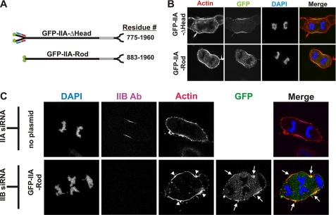
FIGURE 4.
Headless myosin IIA can localize to the furrow in COS-7. A, diagram of GFP-IIA-ΔHead and GFP-IIA-Rod constructs with their corresponding residues. B and C, at 72 h after transfection, COS-7 cells expressing GFP-IIA-ΔHead, or GFP-IIA-Rod or COS-7 cells transfected with MHC IIA siRNA or with MHC IIB siRNA together with GFP-IIA-Rod were fixed and stained for F-actin (red) and for DNA (blue). The samples in panel C were also immunostained for MHC IIB to identify cells that received the siRNA. The images in the right column are merges of all other channels in the panel. Arrows and arrowheads indicate the enrichment of GFP-IIA-Rod and actin, respectively, in furrow zones.

