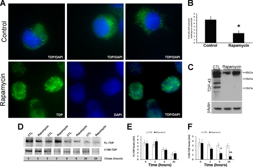
FIGURE 5.
Rapamycin reduces cytosolic TDP-43 immunoreactivity. Cells transfected with C199-TDP were treated with rapamycin or vehicle as described under “Experimental Procedures.” A, representative microphotographs of cells stained with 2E2-D3. Remarkably, rapamycin administration significantly reduced TDP-43 mislocalization as denoted by the increased TDP-43 immunoreactivity (green) in the nucleus (blue). B, quantitative assessment of the number of cells showing cytosolic TDP-43 immunoreactivity after C199-TDP expression. Rapamycin reduced the number of cells showing cytosolic TDP-43 immunoreactivity by ∼60%. p = 0.0016 as determined by t test analysis. C, representative Western blot probed with anti-TDP-43 monoclonal antibody 2E2-D3 showing a significant decrease in the steady-state levels of the C-terminal fragments of TDP-43. D, representative Western blot after pulse-chase experiments. After being pulse-labeled for 2 h, cells were chased for the indicated time points. E and F, quantitative assessment of the FL-TDP and C199-TDP levels after the pulse-chase experiments indicates that the FL-TDP levels decreased similarly in the control (CTL) and rapamycin-treated cells (p > 0.05). In contrast, C199-TDP levels were significantly lower in rapamycin-treated cells compared with control cells at 6 and 24 h, indicating that rapamycin facilitates the turnover of C199-TDP. *, p < 0.01; **, p < 0.001 as determined using one-way analysis of variance, followed by the post hoc Bonferroni test to determine individual differences in groups. DAPI, 4′,6-diamidino-2-phenylindole; AU, arbitrary units.

