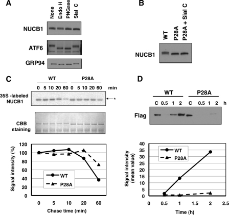
FIGURE 4.
Inefficient export of the P28A mutant from the ER. A, HT1080 cells were lysed in 1% Triton X-100 buffer. The lysates were digested with the indicated glycosidase for 20 h. Each sample was subjected to SDS-PAGE and then underwent immunoblot analysis with anti-NUCB1, anti-ATF6, and anti-KDEL antibodies. B, HT1080 cells were transiently transfected with pNUCB1 or pNUCB1-P28A plasmid (non-tag). The prepared samples, as shown in A, underwent immunoblot analysis with an anti-NUCB1 antibody. C, after transfection with pNUCB1-WT or -P28A (FLAG tag), 293T cells were pulse-labeled for 10 min with [35S]Met/Cys. Then the cells were incubated for the indicated periods in fresh medium to chase 35S-labeled proteins. Each cell lysate was immunopurified with anti-FLAG beads. The samples were subjected to SDS-PAGE, and the gel image was visualized using typhoon (top). The gel was Coomassie Brilliant Blue (CBB)-stained for loading control. Signal intensity was counted using Image J (bottom). D, 293T cells were transiently transfected with the NUCB1-WT or P28A plasmid. After changing to serum-free, fresh medium, each culture medium was collected at the indicated time period. The samples were condensed by an Amicon YM-30 centrifugal ultrafilter and boiled in SDS sample buffer. Then the samples were subjected to SDS-PAGE and immunoblot analysis with an anti-FLAG antibody (top). Signal intensity was counted using Image J (bottom).

