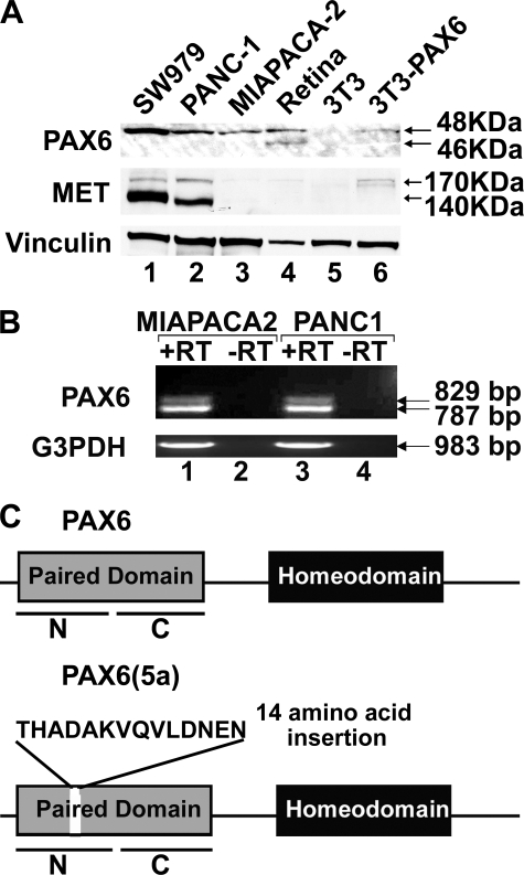
FIGURE 2.
Expression of PAX6 and PAX6(5a) in pancreatic carcinoma cell lines. A, Western analysis of cell lysates. Lysate samples include pancreatic cancer cell lines SW979 (lane 1), PANC-1 (lane 2), and MIAPACA-2 (lane 3), mouse retina (lane 4), PAX6-non-expressing mouse fibroblast cell line 3T3 (lane 5), and 3T3 cells transfected with 3×FLAG-PAX6 (lane 6). Samples are tested for expression of Vinculin (loading control), MET receptor, and PAX6. Expected sizes for PAX6 proteins are 46 kDa (canonical PAX6) and 48 kDa (PAX6(5a) and 3×FLAG-PAX6). Retinal tissues express both forms of PAX6 and are a size control for both isoforms (45). B, reverse transcription-PCR analysis for the expression of PAX6 and PAX6(5a) transcripts in MIAPACA-2 (lanes 1 and 2) and PANC-1 (lanes 3 and 4). The reverse transcription reactions occurs in the presence (lanes 1 and 3) or absence (lanes 2 and 4) of reverse transcriptase. The resulting cDNAs are amplified for primers recognizing glyceraldehyde-3-phosphate dehydrogenase (G3PDH; positive control) or the paired domain of PAX6. The amplicon for canonical PAX6 is 787 bp, and the amplicon for PAX6(5a) is 829 bp. C, schematic of the PAX6 and PAX6(5a) proteins. Proteins are identical (with paired and homeodomain DNA binding domains) except that the PAX6(5a) protein contains a 14-amino acid insertion in the PAI N-terminal subdomain (N) of the paired domain. The paired domain also contains a C-terminal RED subdomain (C).

