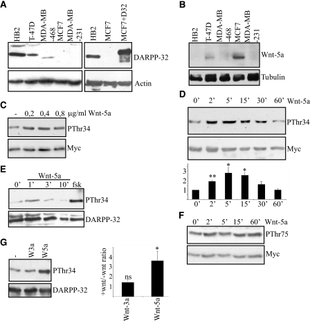
FIGURE 1.
Wnt-5a induces Thr-34 phosphorylation of DARPP-32 in MCF-7 and HB2 breast epithelial cells. A, left panel, expression of DARPP-32 in five different breast epithelial cell lines, detected by anti-DARPP-32 Western blotting. Right panel, comparison of endogenous levels of DARPP-32 in HB2 cells to exogenous levels of Myc-tagged DARPP-32 in transfected MCF-7 cells, detected by anti-DARPP-32 Western blotting. B, expression of Wnt-5a in five different breast epithelial cell lines, detected by an anti-Wnt-5a antibody. C, Western blot showing Thr-34 phosphorylation of DARPP-32 in MCF-7 cells expressing Myc-DARPP-32, after 5 min of stimulation with different concentrations of rWnt-5a, as detected by an anti-phospho-Thr-34-DARPP-32 antibody. D, upper panel, Western blot showing the kinetics of Thr-34-DARPP-32 phosphorylation upon stimulation with 0.4 μg/ml rWnt-5a in DARPP-32-expressing MCF-7 cells. Lower panel, densitometric analysis of three different Western blots. The data are given as means ± S.D. E, Western blot showing Thr-34 phosphorylation of endogenous DARPP-32 induced by Wnt-5a in HB2 cells as detected by anti-phospho Thr-34-DARPP-32 antibody. F, Western blot showing the time kinetics of Thr-75 DARPP-32 phosphorylation upon stimulation with 0.4 μg/ml rWnt-5a in Myc-DARPP-32-expressing MCF-7 cells, anti-phospho Thr-75 DARPP-32 antibody. G, left panel, Thr-34-DARPP-32 phosphorylation after 5 min of stimulation with either 0.4 μg/ml rWnt-5a or 0.1 μg/ml rWnt-3a. Right panel, densitometric analysis of five different Western blots. Wnt-3a or Wnt-5a stimulation was measured relative to no stimulation, which was set to 1. All of the Western blots shown are representative of at least three independent experiments.

