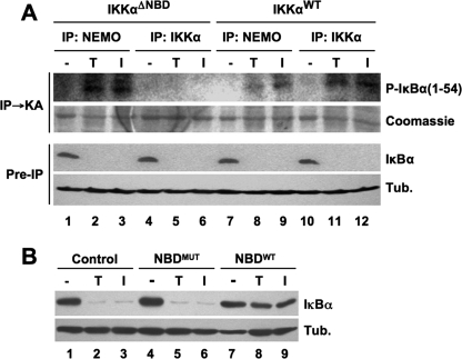
FIGURE 7.
Cytoplasmic IKKαΔNBD complexes are not activated by proinflammatory cytokines. A, IKKαΔNBD and IKKαWT MEFs were either untreated (−) or incubated for 15 min with either TNF (T) or IL-1α (I). Cytoplasmic extracts were prepared, and immunoprecipitations (IP) were performed using anti-NEMO or anti-IKKα as indicated. The precipitated material was used for an immune complex kinase assay (IP → KA) employing glutathione S-transferase-fused IκBα 1–54 as a substrate. Phosphorylated IκBα 1–54 (P-IκBα(1–54)) was detected by autoradiography, and total substrate was visualized by Coomassie staining. Preimmunoprecipitation (Pre-IP) samples from lysates were immunoblotted using anti-IκBα and anti-tubulin (Tub.). B, IKKαΔNBD MEFs were either untreated (Control) or incubated for 15 min with either NBDMUT or NBDWT peptide, as indicated. The cells were then incubated a further 15 min in the absence (−) or presence of either TNF (T) or IL-1α (I). Cytoplasmic lysates were immunoblotted using anti-IκBα and anti-tubulin (Tub.) as shown (right).

