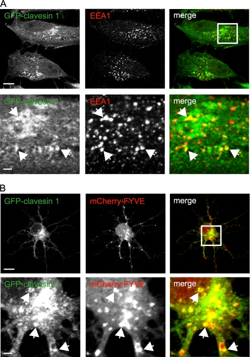
FIGURE 4.
Clavesins localize to endosomes. A, HeLa cells were transfected with plasmid encoding GFP-clavesin 1 and subsequently processed by indirect immunofluorescence with antibody against EEA1 (red). The lower panels are from the area indicated by the white box in the merge image. Bars, 10 and 2 μm for the low and high power images, respectively. B, hippocampal neurons at 6 DIV were transfected with plasmids encoding mCherry-tagged tandem FYVE domains of Hrs (mCherry-FYVE) (red) and GFP-clavesin 1 (green) and were imaged at 7 DIV. The lower panels are from the area indicated by the white box in the merge image. Bars, 10 and 2 μm for the low and high power images, respectively.

