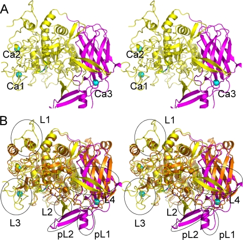
FIGURE 2.
Overall structure of ASP. A, stereo ribbon representation of the overall structure of ASP. The domain architecture is shown in different colors: the N-terminal subtilisin domain (amino acids 3–431) is shown in yellow, and the C-terminal P-domain (amino acids 432–595) is shown in purple. Three bound Ca2+ ions are shown as greenish cyan spheres. B, superimposed stereo view of ASP (yellow and purple as in A) and Kex2 (orange). The four loops in the N-terminal domain of ASP are labeled L1, L2, L3, and L4, whereas the two parts of the unique extra occluding region in the C-terminal domain are labeled as pL1 and pL2.

