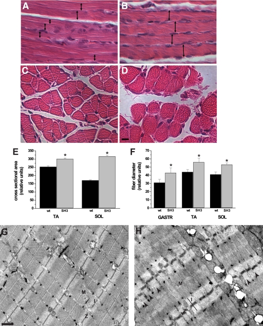
FIGURE 4.
Loss of Bin1 SH3 function results in an unorganized myofibril arrangement. A and B, longitudinal section of hematoxylin/eosin-stained soleus muscle from post-embryonic day 10, wild type, and bin1SH3 mice. The widths of myofibers from the bin1SH3 mice are markedly larger (double-headed arrows). C and D, cross-sectional fiber diameters of the soleus from bin1SH3 mice are also noticeably larger than wild type. Scale bar represents 100 μm. E, cross-sectional area of tibialis anterior (TA) and soleus (SOL) muscles from wild type (wt) and bin1SH3 (SH3) mice were compared. n = 6 muscles per group with at least 60–80 fibers per muscle were counted (*, significantly different, p < 0.001). F, relative fiber diameters of the gastrocnemius (GASTR), tibialis anterior (TA), and soleus (SOL) of p10 wild type (wt) and bin1SH3 (SH3) mice were compared. n = 6 per group (*, significantly different, p < 0.001). G and H, transmission electron microscopy showing the ultrastructure of the gastrocnemius from wild type (G) and bin1SH3 (H) muscle. The M-bands (M), z-disks (Z), and I-bands (I) from bin1SH3 muscle are distended, and the z-disks are periodically misaligned (arrows). Scale bar represents 500 nm.

